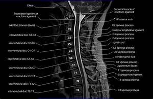
Medical imaging is a cornerstone of modern healthcare, enabling clinicians to make accurate diagnoses and devise effective treatment plans. In the realm of head and neck pathology, computed tomography (CT) has proven to be indispensable. The ability to visualize both hard and soft tissue in high detail allows for the early detection and management of a wide range of medical conditions. One of the most crucial aspects of CT imaging is the ability to use sagittal labeling to map key anatomical structures in the head and neck region. This technique provides a side view of these structures, enhancing the understanding of their spatial relationships and ensuring precise diagnosis. In some cases, Ct Head And Neck Sagittal Labeling has been the difference between life and death. This article explores several case studies where sagittal labeling played a critical role in saving lives.
Case Study 1: Trauma and Cervical Spine Injury
Background
A 45-year-old male was admitted to the emergency department after a high-speed motor vehicle collision. Upon arrival, the patient was conscious but complaining of severe neck pain and difficulty moving his limbs. Initial physical examination raised concern for a cervical spine injury, and a CT scan was ordered to assess the extent of the damage. The sagittal view of the CT scan would prove to be pivotal in diagnosing the injury.
The Role of Sagittal Labeling
The CT images revealed significant disruption in the alignment of the cervical spine, particularly between the C5 and C6 vertebrae. Sagittal labeling was used to mark the exact location of the fracture and displacement. The sagittal view offered a clear picture of the relationship between the fractured vertebrae and the spinal cord, which was at risk of compression due to the misalignment.
By accurately labeling the key anatomical structures—including the vertebrae, spinal cord, and surrounding muscles—the medical team could see that the displaced bone fragments were dangerously close to the spinal cord. This information was crucial for determining the severity of the injury and the appropriate intervention.
Outcome
Due to the precise sagittal labeling, the surgical team was able to quickly assess the need for immediate spinal stabilization surgery. The operation was performed within hours, and the patient’s spinal cord was spared from permanent damage. The patient was eventually able to regain full mobility, thanks to the timely intervention that was informed by the sagittal CT scan.
Case Study 2: Detection of Head and Neck Cancer
Background
A 60-year-old female presented with a persistent sore throat, difficulty swallowing, and unexplained weight loss. After an initial evaluation, a CT scan of the head and neck was ordered to rule out potential malignancies. Given the patient’s symptoms and medical history, the clinical team suspected that a tumor might be present. Sagittal CT imaging, coupled with sagittal labeling, would reveal critical information.
The Role of Sagittal Labeling
The CT scan revealed an unusual mass in the left side of the neck, extending from the pharynx down to the upper mediastinum. Sagittal labeling of the CT images highlighted the precise location of the mass, which appeared to involve not only the lymph nodes but also the surrounding soft tissues and blood vessels.
By accurately labeling the anatomical landmarks, including the carotid artery and jugular vein, the team could clearly see how the tumor was infiltrating these vital structures. This sagittal view provided invaluable information regarding the tumor’s proximity to critical structures, such as the airway and major vascular networks.
Outcome
Thanks to the sagittal labeling of the CT images, the multidisciplinary oncology team was able to stage the cancer with precision and make a well-informed decision regarding treatment. Surgical resection was planned with careful consideration of the tumor’s location, and postoperative radiation therapy was recommended to target any remaining cancer cells. The patient underwent surgery successfully, and the tumor was removed with clear margins. The early detection made possible by the sagittal CT scan significantly improved the patient’s prognosis, and she has remained cancer-free for several years post-treatment.
Case Study 3: Carotid Artery Dissection
Background
A 35-year-old male experienced sudden onset of severe, sharp pain in his neck, along with dizziness and blurred vision. After initial evaluation, the patient was suspected of having a vascular event, and a CT angiogram of the head and neck was ordered. Sagittal CT images would provide essential insights into the diagnosis.
The Role of Sagittal Labeling
The CT scan revealed a dissection of the right carotid artery, a potentially life-threatening condition. The sagittal view of the CT images was instrumental in visualizing the tear in the artery’s wall and its impact on blood flow. Sagittal labeling allowed the radiologist to mark the precise location of the dissection and identify any areas where blood flow had been compromised.
By labeling key anatomical structures such as the carotid artery, jugular vein, and the vertebral artery, the team could evaluate the extent of the dissection and its potential to cause a stroke. The sagittal labeling also revealed a close relationship between the dissection and the vertebral artery, a critical vessel supplying blood to the brainstem.
Outcome
The sagittal CT images guided the decision to initiate anticoagulation therapy immediately, preventing further clot formation and reducing the risk of a stroke. The patient was closely monitored, and the dissection healed without further complications. The timely recognition of the carotid artery dissection, made possible by the detailed sagittal CT labeling, ultimately prevented a life-threatening stroke and ensured a favorable outcome.
Case Study 4: Nasopharyngeal Obstruction
Background
A 5-year-old child was brought to the emergency department with signs of respiratory distress, including stridor and difficulty breathing. Initial clinical suspicion was that the child had an upper airway obstruction, possibly from a foreign body or a mass. A CT scan of the head and neck was ordered to evaluate the cause of the obstruction, and sagittal labeling would again prove invaluable in determining the diagnosis.
The Role of Sagittal Labeling
The CT images revealed a large mass located in the nasopharynx, which was blocking the airway and causing significant narrowing of the air passages. Sagittal labeling was critical in identifying the size and location of the mass, which appeared to be a benign tumor, such as a juvenile nasopharyngeal angiofibroma.
The sagittal view allowed clinicians to assess how the mass was impinging on the airway and surrounding structures, such as the Eustachian tube and the carotid artery. Labeling the key anatomical structures allowed the surgical team to plan the best approach for removing the mass while avoiding damage to the nearby critical structures.
Outcome
The child underwent surgery to remove the nasopharyngeal mass, with the operation carefully guided by the labeled CT images. Postoperative recovery was smooth, and the child was able to breathe normally following the procedure. The early identification of the obstruction through sagittal labeling was instrumental in preventing life-threatening respiratory distress and ensuring a successful surgical outcome.
Conclusion
CT head and neck sagittal labeling has proven to be a life-saving technique in various clinical scenarios, from traumatic injuries to complex pathologies like cancer and vascular abnormalities. These case studies demonstrate how precise sagittal imaging, coupled with accurate labeling, can provide critical insights into the anatomical relationships within the head and neck region, enabling clinicians to make timely and informed decisions. In each case, sagittal CT labeling helped guide diagnosis, treatment planning, and surgical intervention, ultimately saving lives and improving patient outcomes. As imaging technology continues to evolve, the importance of mastering techniques like sagittal labeling will only grow, ensuring that medical professionals can continue to provide the highest level of care. Visit Health Dady to get more information.


